How to Read Echo Bubble Study Test Results
Written by Dr. Ahmed Ali and Dr. Alexander Nossikoff
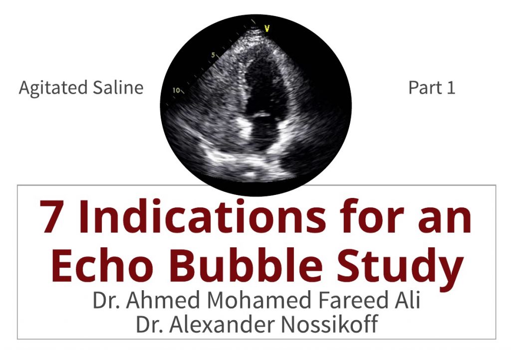
Agitated saline "Echo Bubble written report" is frequently a neglected complementary echocardiographic technique. What is an echo bubble study? What are the indications and interpretation for the use of agitated saline, "bubble study"? This week Dr. Ali and Dr. Nossikoff will provide us with seven indications for an repeat chimera report and review how to translate the findings. Read more than about both doctors in this weeks educator spotlight articles.
Echo Chimera Study: What is it?
An Echo Chimera Written report is an injection of saline after agitation with air to create micro-bubbles that are ultrasound reflective into a vein in lodge to reach and opacify the correct heart chambers, the coronary sinus in cases of persistent left superior vena cava (PLSVC), or the pericardium during pericardiocentesis.
7 Indications for an Repeat Bubble Written report
1- Detection of shunts (PFO, ASD, pulmonary)
2- Detection of persistent left superior vena cava.
3- Intensifying TR signal when you lot accept difficult estimating RV systolic pressure
iv- Delineating right heart borders and masses (including RV wall thickness).
five- Improving imaging of the pulmonary trunk and arteries, especially when looking for thrombi, which will appear as contrast filling defects.
6- During echo-guided pericardiocentesis.
vii- For central venous line control afterwards insertion.
How to Perform and Interpret Findings
one. Detection of Shunts
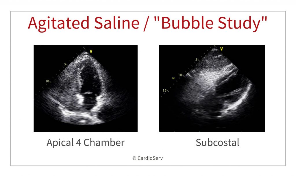
In general; the advent of micro-bubbling on the left side of the middle afterwards their appearance in the right heart chambers is considered positive shunt report.
Most commonly, apical four-bedroom view is used, likewise parasternal short-axis view at the level of the atrial septum (aortic valve level) or a subcostal four-chamber view may exist used.
ASD / PFO
In example of ASD or PFO; the bubbles should announced on the left side of the heart inside the start three to 4 cardiac cycles.
If no bubbles appear in the left side and so the patient is instructed to perform Valsalva maneuver (which leads to opening of PFO during it's release) and bubbles appear in the left side in case of PFO. Failure to demonstrate transient leftward bowing atrial septum with Valsalva release indicates insufficient operation of the maneuver and should be repeated.
During TEE – patients are commonly not able to perform proper Valsalva. Most operators now use external liver compression followed by release causing sudden increase in IVC flow towards RA with similar effects equally Valsalva release. Some operators utilise cough likewise.
Some other finding is the so called "negative jet" which appear every bit filling defect within the fully opacified right side of the heart in front end of ASD due to left to right shunting. This could exist mistaken with the competitive catamenia from inferior vena cava along the RA side of the atrial septum.
Pulmonary Arteriovenous Malformations (PAVM's)
In case of pulmonary arteriovenous malformations (PAVM's) which are abnormal dilated vessels provide a correct-to-left shunt between the pulmonary and systemic circulation and the shunt is actress cardiac; the micro-bubbling may announced in the left side of the center afterwards five or more cardiac cycles "late bubbles". In hepatopulmonary syndrome, which is typically characterized past unexplained desaturation in a cirrhotic patient, the PAVMs are microscopic and will not be visible on CTPA, right-to-left shunting appearance on echocardiography volition be the same as in archetype PAVMs every bit unremarkably seen in Osler-Weber-Rendu syndrome. Agitated saline can be used every bit zippo radiation technique for screening of family members of patients with Osler-Weber-Rendu syndrome.
Keep in mind in some patients with large RA and irksome filling due to loftier RA pressures, complete RA opacification will take longer and chimera appearance in LA may be delayed mimicking shunting at lung level.
2. Persistent Left Superior Vena Cava
In case of dilated coronary sinus and doubtful persistent left superior vena cava; the cannula must exist inserted in the left arm, and the agitated saline should exist injected through the left arm.
The test is positive for LSVC if the agitated saline appears in the coronary sinus before appearance in the right side of the eye.
Imaging is ameliorate through the parasternal long centrality view. G-mode can be employed for better temporal resolution, with the beam centered on CS and RVOT.
Experienced operators use angulated A4C with posterior tilt showing CS opening into RA. As more than complex anatomic variants be, iind injection into right arm is suggested.
iii. Intensifying TR Signal
In instance that the TR bespeak past CW is suboptimal and pulmonary artery systolic pressure is important for clinical decision making; injecting agitating saline and obtaining CW of TR brand the signal more visible and measurable but CW gain should be decreased equally agitated saline brand cause some noise. In this case gelofusine will cause a lot of background noise and should exist better exist avoided.
iv. Delineate Correct Heart Borders and Masses
Sometimes in suboptimal views; the RV borders are not clear, or you may have doubt about a mass or trabeculations within the RV cavity. Injecting agitated saline may help delineating RV borders for accurate measurements as it goes inside the myocardial recesses and split up the dense compacted myocardium from the cavity. This also helps to delineate RV free wall.
5. Improved Imaging of Pulmonary Arteries
For proper conclusion of pulmonary avenue size & sub/supravalvular area; agitated saline may assist if the image is suboptimal. Thrombi and masses tin can appear equally filling defects.
6. During Echo Guided Pericardiocentesis
During pericardiocentesis; agitated saline could exist used during the procedure to differentiate if puncture is within the pericardium or in 1 the cardiac chambers (mostly RV) by noticing the microbubbles either in the pericardium or within a chamber.
seven. Key Venous Line Control after Insertion
Contrast should appear immediately in RA subsequently forceful push through 1 of the central line ports, if not actualization at all this means arterial cannulation, if appearing late – this means coiling of the catheter. When using a central venous line port and forceful saline push button some agitation takes place fifty-fifty without air, then you can skip the step with 0.five – 1 ml of air.
Summary
Thank you to both Dr. Ahmed Mohamed Fareed Ali and Dr. Alexander Nossikoff for breaking downwardly 7 indications for an echo bubble study in the first part of our 2-part web log serial on agitated saline. Next week both doctors will explicate how to perform the procedure, what supplies are needed and tips for optimizing imaging.

Dr. Alexander Nossikoff
References
Gupta, Southward. K., Shetkar, S. S., & Ramakrishnan, S. (2015). Saline Dissimilarity Echocardiography in the Era of Multimodality Imaging — Importance of " Bubbling Information technology Correct ." Echocardiography, 1707–1719. https://doi.org/10.1111/echo.13035
Marriott, One thousand., Ultrasound, M. C., Manins, Five., Forshaw, A., Ultrasound, Thou. C., Wright, J., & Pascoe, R. (2013). Detection of Right-to-Left Atrial Communication Using Agitated Saline Contrast Imaging : Experience with 1162 Patients and Recommendations for Echocardiography. Journal of the American Society of Echocardiography, 26(1), 96–102. https://doi.org/10.1016/j.echo.2012.09.007
Porter, T. R., Abdelmoneim, S., Belcik, J. T., Mcculloch, M. L., Mulvagh, S. L., Olson, J. J., … Wei, K. (2014). Guidelines for the Cardiac Sonographer in the Performance of Contrast Echocardiography : A Focused Update from the American Society of Echocardiography. Journal of the American Society of Echocardiography, 27(8), 797–810. https://doi.org/10.1016/j.echo.2014.05.011
Puledda, F., Toscano, Chiliad., Pieroni, A., Veneroso, G., Piero, 5. Di, & Vicenzini, E. (2016). Right-to-left shunt detection sensitivity with air – saline and air – succinil gelatin transcranial Doppler. International Periodical of Stroke, 11(2), 229–238. https://doi.org/ten.1177/1747493015609938
Romero, R., Frey, J. L., Schwamm, L. H., Demaerschalk, B. G., Chaliki, H. P., Parikh, G., … Babikian, 5. L. (2009). Cognitive Ischemic Events Associated With ' Bubble Study ' for Identification of Right to Left Shunts. Stroke, 2342–2348. https://doi.org/10.1161/STROKEAHA.109.549683
Source: https://www.cardioserv.net/indications-echo-bubble-study/
0 Response to "How to Read Echo Bubble Study Test Results"
Enviar um comentário